For the past 40 years, disease progression has been evaluated through lung function tests and plain chest radiographs. The implementation of multi-detector chest CT scan technology has provided clinicians with a more sensitive method for imaging CF lung damage. Various types of imaging techniques are now used to determine the presence and extent of lung disease in patients with CF, including CT and chest magnetic resonance imaging (MRI). In addition, scoring systems have been developed to quantify and characterize the structural abnormalities detected throughCTandMRI in patients withCFat various stages of the disease. These systems assess structural changes such as bronchiectasis, trapped air, airway wall thickening, mucus, and opacities. More recently, chest MRI techniques have been developed allowing the assessment of functional characteristics of the lung, as well as the evaluation of lung morphology. Positron emission tomography (PET) imaging with [18F]fluorodeoxyglucose ([18]FDG; FDG-PET) can also be used as a non-invasive technique to quantify lung inflammation.
The prevalence of pancreatic fatty replacement in cystic fibrosis varies with age and is most frequently identified in older patients. In an autopsy series of 27 cases of cystic fibrosis that spanned a period of 5 years, diffuse fatty replacement was reported in 15 cases (56%) . The mean age of patients with fatty replacement was 17 years, whereas the mean age of patients without fatty replacement was only 11 years. At MR imaging, the reported prevalence of diffuse fatty change varies from 51% to 75% , with partial fatty replacement seen in 7%–29% of cases and pancreatic atrophy in 27%–35%. They reported pancreatic enlargement in association with fatty change at MR imaging in nine of 17 adult patients (53%) with cystic fibrosis. Our experience has shown that it can be difficult to distinguish the fat-replaced pancreas from normal retroperitoneal fat, making it difficult to assess the size of the gland. However, diffuse pancreatic enlargement was apparent in five of 34 patients (15%) with cystic fibrosis who had undergone pancreatic MR imaging at our institution. Fatty infiltration of the pancreas can also be readily identified at computed tomography, but this modality involves the use of ionizing radiation. Typical ultrasonographic (US) appearances, including increased parenchymal echogenicity and pancreatic atrophy, have also been described by several authors , although MR imaging is superior to US in demonstrating fatty infiltration .
Sources:
http://www.appliedradiology.com/articles/imaging-of-the-pancreas-part-1
http://pubs.rsna.org/doi/full/10.1148/radiographics.20.3.g00ma08767
Pediatric Pulmonology 50:302–315 (2015)
Novel Outcome Measures for Clinical Trials in Cystic Fibrosis
Harm A.W.M. Tiddens, Michael Puderbach, Jose G. Venegas,
Felix Ratjen, Scott H. Donaldson, Stephanie D. Davis,
Steven M. Rowe, Scott D. Sagel, MD, Mark Higgins, and
David A. Waltz,
samedi 16 mai 2015
jeudi 19 mars 2015
Quick introduction to imaging
http://www.nhlbi.nih.gov/health/health-topics/topics/cf/signs |
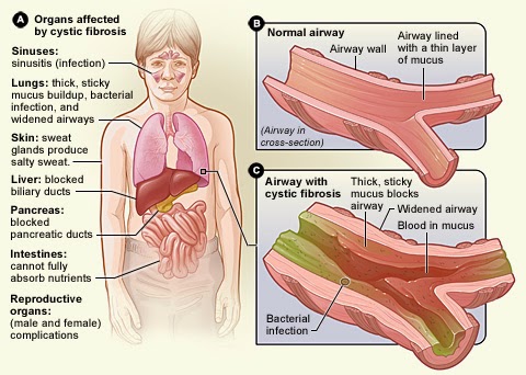
As we said the cystic fibrosis affect not only the lungs, as we can see on the picture it's affect lots of organs.
The signs and symptoms of cystic fibrosis (CF) vary from person to person and over time. Sometimes you'll have few symptoms. Other times, your symptoms may become more severe.
One of the first signs of CF that parents may notice is that their baby's skin tastes salty when kissed, or the baby doesn't pass stool when first born.
Most of the other signs and symptoms of CF happen later. They're related to how CF affects the respiratory, digestive, or reproductive systems of the body.
To diagnose and follow a patient we need to see what is appening in those organs that's why imaging is so important and why we will introduce it to you. The following image is a magnetic resonance imaging of lungs (MRI). MRI is often use to follow patient to see if the mucus density is too hight or not.
Source:https://www.aerzteblatt.de/int/archive/article?id=156541&src=search MRI in cystic fibrosis. A 7-year-old girl in long-term treatment for cystic fibrosis at a pediatric pneumology center. a) Chest radiograph. b) MRI on the same day; the patient was sedated but free breathing. The T1-weighted contrast studies show clear bronchial wall thickening and varicose bronchiectasis, most pronounced in the upper lobes (arrows), and reduced signal apically compared to basally. |
Inscription à :
Articles (Atom)