For the past 40 years, disease progression has been evaluated through lung function tests and plain chest radiographs. The implementation of multi-detector chest CT scan technology has provided clinicians with a more sensitive method for imaging CF lung damage. Various types of imaging techniques are now used to determine the presence and extent of lung disease in patients with CF, including CT and chest magnetic resonance imaging (MRI). In addition, scoring systems have been developed to quantify and characterize the structural abnormalities detected throughCTandMRI in patients withCFat various stages of the disease. These systems assess structural changes such as bronchiectasis, trapped air, airway wall thickening, mucus, and opacities. More recently, chest MRI techniques have been developed allowing the assessment of functional characteristics of the lung, as well as the evaluation of lung morphology. Positron emission tomography (PET) imaging with [18F]fluorodeoxyglucose ([18]FDG; FDG-PET) can also be used as a non-invasive technique to quantify lung inflammation.
The prevalence of pancreatic fatty replacement in cystic fibrosis varies with age and is most frequently identified in older patients. In an autopsy series of 27 cases of cystic fibrosis that spanned a period of 5 years, diffuse fatty replacement was reported in 15 cases (56%) . The mean age of patients with fatty replacement was 17 years, whereas the mean age of patients without fatty replacement was only 11 years. At MR imaging, the reported prevalence of diffuse fatty change varies from 51% to 75% , with partial fatty replacement seen in 7%–29% of cases and pancreatic atrophy in 27%–35%. They reported pancreatic enlargement in association with fatty change at MR imaging in nine of 17 adult patients (53%) with cystic fibrosis. Our experience has shown that it can be difficult to distinguish the fat-replaced pancreas from normal retroperitoneal fat, making it difficult to assess the size of the gland. However, diffuse pancreatic enlargement was apparent in five of 34 patients (15%) with cystic fibrosis who had undergone pancreatic MR imaging at our institution. Fatty infiltration of the pancreas can also be readily identified at computed tomography, but this modality involves the use of ionizing radiation. Typical ultrasonographic (US) appearances, including increased parenchymal echogenicity and pancreatic atrophy, have also been described by several authors , although MR imaging is superior to US in demonstrating fatty infiltration .
Sources:
http://www.appliedradiology.com/articles/imaging-of-the-pancreas-part-1
http://pubs.rsna.org/doi/full/10.1148/radiographics.20.3.g00ma08767
Pediatric Pulmonology 50:302–315 (2015)
Novel Outcome Measures for Clinical Trials in Cystic Fibrosis
Harm A.W.M. Tiddens, Michael Puderbach, Jose G. Venegas,
Felix Ratjen, Scott H. Donaldson, Stephanie D. Davis,
Steven M. Rowe, Scott D. Sagel, MD, Mark Higgins, and
David A. Waltz,
Bio cell : Transporters
samedi 16 mai 2015
jeudi 19 mars 2015
Quick introduction to imaging
http://www.nhlbi.nih.gov/health/health-topics/topics/cf/signs |
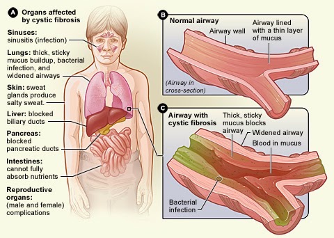
As we said the cystic fibrosis affect not only the lungs, as we can see on the picture it's affect lots of organs.
The signs and symptoms of cystic fibrosis (CF) vary from person to person and over time. Sometimes you'll have few symptoms. Other times, your symptoms may become more severe.
One of the first signs of CF that parents may notice is that their baby's skin tastes salty when kissed, or the baby doesn't pass stool when first born.
Most of the other signs and symptoms of CF happen later. They're related to how CF affects the respiratory, digestive, or reproductive systems of the body.
To diagnose and follow a patient we need to see what is appening in those organs that's why imaging is so important and why we will introduce it to you. The following image is a magnetic resonance imaging of lungs (MRI). MRI is often use to follow patient to see if the mucus density is too hight or not.
Source:https://www.aerzteblatt.de/int/archive/article?id=156541&src=search MRI in cystic fibrosis. A 7-year-old girl in long-term treatment for cystic fibrosis at a pediatric pneumology center. a) Chest radiograph. b) MRI on the same day; the patient was sedated but free breathing. The T1-weighted contrast studies show clear bronchial wall thickening and varicose bronchiectasis, most pronounced in the upper lobes (arrows), and reduced signal apically compared to basally. |
mercredi 17 décembre 2014
Cystic fibrosis: Explanations
Description of one genetic disease
The cystic fibrosis, also known as mucoviscidosis, is a genetic disease that affects mostly lungs but also the pancreas, liver, and intestine. Cystic fibrosis is due to a mutation of only one gene which is the one who help to produce CFTR (cystic
fibrosis transmembrane conductance regulator), a transmembrane protein that help Cl- and Na+ to pass through the membrane.
Where can we found it?
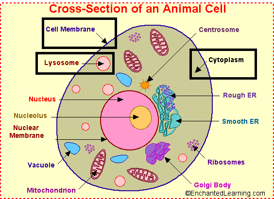
As we can see on the picture we can find these in the cell membrane, in lysosome and in the cytoplasm, in the early and late endosome.
http://www.enchantedlearning.com/subjects/animals/cell/ |
How does a protein problem lead to so much trouble?
http://healthylookout.com/?p=1145 |
On image 2, it's a CFTR produced by a mutation: As we can see, the canal is closed and the result of a lack of NaCl in the mucus is a very sticky mucus that cover all the epithelium. Then the accumulation of this mucus causes respirator deficiency and infections.
jeudi 29 mai 2014
Co-transporters:
I) Historical:
In august 1960 in Prague, the Czech Republic Robert K.Crane discovered the mecanism of the sodium-glucose cotransport for the glucose intestinal absorption. It was the most important concerning later the carbohydrates absorption in the 20th century.
II) What are these proteins?
Co-transporters, or coupled transporters, are a subcategory of transporters that couple the favorable movement of one molecule with its concentration gradient and unfavorable movement of another molecule against its concentration gradient.
According to the path of these two elements, we'll talk about symporters (ion and molécule travel the membrane in the same direction) or antiporters (these two chemical species travel the membrane in opposite directions).
http://ressources.unisciel.fr/biocell/chap2/co/module_Chap2_2.html |
Uniporters:
mardi 17 décembre 2013
Pumps Na+/K+ ATPase:
History:
Na+/K+-ATPase was discovered by Jens Christian Skou in 1957 while working as assistant professor at the Department of Physiology, University of Aarhus, Denmark. He published his work that year.In 1997, he received one-half of the Nobel Prize in Chemistry "for the first discovery of an ion-transporting enzyme, Na+/ K+-ATPase."
Function:
Export of sodium from the cell provides the driving force for several secondary active transporters , which import glucose, amino acids, and other nutrients into the cell by use of the sodium gradient.
Failure of the Na+-K+ pumps can result in swelling of the cell. A cell's osmolarity is the sum of the concentrations of the various ion species and many proteins and other organic compounds inside the cell. When this is higher than the osmolarity outside of the cell, water flows into the cell through osmosis. This can cause the cell to swell up and die (the lyse). The Na+-K+
pump helps to maintain the right concentrations of ions. Furthermore,
when the cell begins to swell, this automatically activates the Na+-K+ pump.
http://www.asso-etud.unige.ch/aecb/articles/notes_cours/1ere/biolobarja2011.pdf |
lundi 16 décembre 2013
Aquaporins:
1) Historical:
These aquaporins have been discovered in 1992 by an american biologist named Peter Agre during a studying about red blood cells: it was during an experience about an injection of a "copy" of DNA (or RNAm) of a protein in an amphibian's egg. In 2003, the biologist was rewarded by the Nobel Prize of Chemistry.
2) What is the aquaporin?
At the human's body, aquaporins are really important for the operation of the kidneys. Cellulars have a plasma membrane which no accept the pass of the water. However, they have aquaporins which can pass the water easier through the cellular. In fact, thanks to hydrogen bonds (chemestry bond between oxygen atoms and hydrogen atoms) and proteins' charges, that the water travel in the plasma membrane.
Inscription à :
Articles (Atom)